EVENT / 1961 / BIOLOGY
The Experiments That Made Stem Cells Measurable
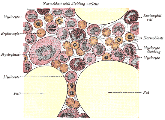
FIG. 1 · BEFORE 1858 · PUBLIC DOMAIN
Safety at a Glance
Cell therapies
- Main risk
- Unapproved products: FDA reports of blindness, tumours and infections [FDA-S01]
- Critical warning
- A trial listing or FDA registration does not mean a product is approved [FDA-S01]
- Lethal dose
- No universal threshold in the reviewed sources; harms depend on procedure, product, donor and patient [CARE-S01]
A rescue that needed explaining
Around the turn of the 1960s, two scientists at the Ontario Cancer Institute in Toronto, Ernest McCulloch and James Till, set out to understand how radiation damages living cells. The question was urgent for several reasons, according to the Lasker Foundation's account of their work. Doctors wanted to know when radiation therapy defeated cancer. The Cold War raised fears of soldiers and civilians exposed to nuclear weapons. And a new treatment, bone marrow transplantation, was showing that fresh marrow could rescue an animal whose own blood-making system had been destroyed HIST-S01.
The identity and properties of the marrow cells responsible still needed to be established. Blood cells were familiar enough: red cells that carry oxygen, white cells that fight infection, platelets that stop bleeding. Physicians could recognise immature forms of them in the marrow. The researchers sought testable evidence of the cells that sustained that production HIST-S01.
Till and McCulloch's experiments in mice, published from 1960 onward, did not solve that mystery in a single stroke. They developed a way to count cells by their capacity to form colonies. That shift, from a hypothesis to a measurable object, is the real milestone of 1961.
The idea before the evidence
The notion of a "stem cell" was not new in 1961. By the turn of the twentieth century, the Lasker account notes, scientists were already proposing that the body contained self-renewing cells that could give rise to many specialised kinds HIST-S01. What was missing was a way to show that such cells actually existed, and to study them.
Today, the U.S. National Institutes of Health describes stem cells by two defining abilities BIO-S01:
- Self-renewal: a stem cell can make more stem cells, so the supply does not run out.
- Differentiation: a stem cell can give rise to specialised cells, such as the different types of blood cell.
Different stem cells vary in potency, meaning how many kinds of cell they can produce. "Adult" stem cells, found in particular tissues, generally produce the specialised cells of their own tissue or organ. Pluripotent stem cells, which include embryonic stem cells and cells reprogrammed in the laboratory, can give rise to all the cell types of the adult body BIO-S01. The blood-forming stem cells at the centre of this story belong firmly in the first group. They make blood, not hearts or brains.
Counting what you cannot see
Till and McCulloch's first step was to measure how sensitive marrow cells were to radiation. They gave mice enough radiation to destroy their ability to make blood, then injected marrow from unirradiated donors that had itself been exposed to varying amounts of radiation. By seeing how much marrow was needed to save the animals, they could estimate how many marrow cells survived each dose HIST-S01.
Then came the observation that made their name. When the researchers examined the mice some days after transplantation, they found small lumps, or nodules, on the spleen. The nodules contained dividing cells, and some of those cells were developing into the three main types of blood cell. Crucially, the number of nodules rose in direct proportion to the number of living marrow cells injected. The cells that formed them were rare HIST-S01.
The pattern reminded the researchers of a standard technique in microbiology. Spread bacteria on a dish, and each living bacterium multiplies into a visible colony; count the colonies, and you know how many living cells you started with. In the same way, counting spleen nodules, which they came to call "colonies," gave a functional measure of the cells in marrow capable of producing them HIST-S01. This "spleen colony" assay, reported in 1961 HIST-S02, was a counting tool. It measured these cells by what they did, rather than by identifying a purified population from appearance alone HIST-S01.
The limits matter as much as the achievement. The assay worked in mice, not people. It did not purify the cells or produce a picture of them. And on its own, it could not answer an obvious objection: was each colony grown from a single cell, or from a clump of several different cells that happened to land together HIST-S01?
One cell or many? Becker's markers
Settling that question required a way to tag individual cells. The team realised that radiation itself could supply the tags: at low frequency, it produces cells with visibly abnormal chromosomes, and those abnormalities are passed on when the cells divide. If dividing cells in a colony shared the same rare chromosomal mark, that would support their descent from one marked ancestor HIST-S01.
The person who did the painstaking work was Andrew Becker, a graduate student working with McCulloch and Till. Examining cells from many spleen colonies, he found a few colonies containing cells with distinctive chromosomes, and in each of those, almost all the dividing cells carried the same unique alteration HIST-S01. Becker, McCulloch and Till published the result in Nature in 1963. It was evidence that the colonies were clones: groups of cells descended from a single cell HIST-S01.
That is a separate result from the 1961 assay. The assay showed that something in the marrow could be counted. Becker’s markers supplied evidence for single-cell origins in the marked colonies.
Making more of themselves: Siminovitch and self-renewal
A cell that could produce blood but not replace itself would eventually run out. So the next question was whether colony-forming cells could renew themselves. With their colleague Louis Siminovitch, Till and McCulloch broke up individual spleen colonies and injected the cells into a fresh set of irradiated mice. If the colonies contained new colony-forming cells, the second mice would grow colonies of their own. They did HIST-S01. Siminovitch, McCulloch and Till reported the work in 1963 HIST-S01.
The experiments also revealed something unexpected. Some colonies held many new colony-forming cells, others few or none, and the variation did not seem to be inherited. Till developed a theory in which chance helped decide whether a stem cell would divide into two new stem cells or start to specialise. He tested the model with computer simulation and published it with McCulloch and Siminovitch in 1964 HIST-S01. That model describes variability in stem-cell behaviour. It does not mean that nothing controls what stem cells do.
Further work with mutant mouse strains in the mid-1960s pointed to both sides of that control. In one anaemic strain, the defect lay in the blood-forming stem cells themselves. In another, the stem cells were normal, but the surrounding tissue failed to support them. That showed the importance of the cell's environment, and it helped set the stage for discovering the signalling proteins that guide blood-cell development HIST-S01.
By the early 1970s, in the Lasker account's summary, the team's work showed that marrow transplants owe their restorative power to cells that can divide, renew themselves and produce all three main types of blood cell HIST-S01. Self-renewal and the capacity to produce specialized descendants remain central to the stem-cell concept. One caution: the cells that formed spleen colonies in these experiments should not be assumed identical to every population that modern researchers call blood stem cells.
FIG. 2 · 2018 · CC BY-SA 4.0
What the experiments made possible
The most direct legacy of this work is in the clinic. Today the U.S. National Cancer Institute describes stem cell transplants as procedures that restore blood-forming stem cells in people whose own have been destroyed by the high doses of chemotherapy or radiation used to treat certain cancers and other disorders. The transplanted cells travel to the bone marrow and take the place of those destroyed CARE-S01. They can come from the patient (an autologous transplant) or from a donor (allogeneic), and from bone marrow, circulating blood or umbilical-cord blood CARE-S01.
In donor transplants, the new immune cells can also help in another way. NCI describes a graft-versus-tumour effect, in which the donor's white blood cells attack cancer cells that remain after treatment CARE-S01. Till and McCulloch's work, in the Lasker Foundation's words, "explained the basis" of bone marrow transplantation HIST-S01. They did not invent the procedure, which was already being explored, but their experiments helped explain why it worked.
These transplants are a treatment, not an enhancement. They replace a system that has been destroyed. They are also hard on the body, as the safety section below explains.
From repair to "regeneration"
Stem-cell research did not stop with blood. In 1962, the year after the spleen colony assay, John Gurdon showed that a nucleus taken from a mature frog intestinal cell could direct an egg to develop into a normal tadpole. Specialisation, it turned out, could be reversed. In 2006, Shinya Yamanaka showed that mature cells from mice could be reprogrammed into pluripotent stem cells by introducing a small number of genes. Gurdon and Yamanaka shared the 2012 Nobel Prize in Physiology or Medicine LATER-S01.
That later work belongs to a different branch of the story. Reprogrammed and embryonic cells are pluripotent; the blood-forming cells Till and McCulloch studied are not. The Toronto experiments helped establish that stem cells could be measured and studied rigorously in adult animals, and the Lasker account credits them with encouraging the search for other stem cells, including embryonic ones HIST-S01. But there is no simple straight line from a mouse spleen in 1961 to a reprogrammed cell in 2006.
Till and McCulloch received the 2005 Albert Lasker Basic Medical Research Award for their work HIST-S01. Accepting it, McCulloch noted that the breadth of research worldwide helps society decide "when it is appropriate to apply new knowledge in the clinical setting" HIST-S01. It is a modest statement, and a useful one.
The gap between science and sales
For a history of human enhancement, stem cells are tantalising. A cell that can renew itself and rebuild tissue sounds like a route not just to repair but to rejuvenation. FDA warnings reveal a commercial market making claims that extend beyond established treatments.
The U.S. Food and Drug Administration warns that unapproved "regenerative medicine" products, including stem cells, fat-derived cells, umbilical-cord and amniotic products, and exosomes, continue to be marketed broadly for a wide range of diseases and conditions. It says it has received reports of blindness, tumour formation, infections and other harms linked to their use FDA-S01. It cautions that a listing on the clinical-trials registry, or a company's registration with the FDA, is often used to suggest legitimacy, but does not mean a product is approved FDA-S01.
At the same time, some cell-based products are licensed. The FDA's current list of approved cellular and gene therapy products includes licensed cord-blood products and others, such as RYONCIL, which is indicated for a specific, serious complication of donor transplants in children: steroid-refractory acute graft-versus-host disease FDA-S02; FDA-S03. Approval of that kind is for a defined product and use. It is not an endorsement of stem cells in general.
That is the honest shape of the field 65 years after the spleen colony assay. Blood-stem-cell transplants are an established, life-sustaining treatment for specific conditions. Other cell products have specific approvals. The FDA also warns about widely marketed unapproved offerings. Till and McCulloch's achievement was to make stem cells something scientists could measure. The work of measuring what stem-cell treatments actually do, and for whom, continues.
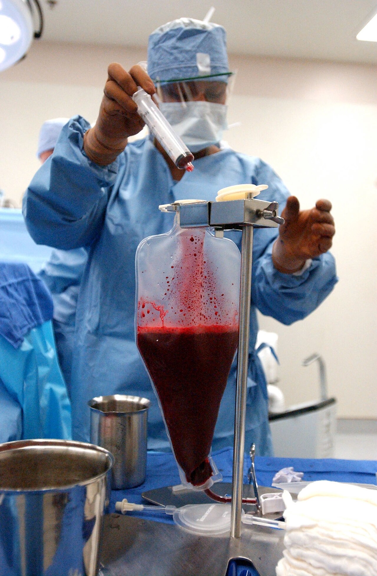
FIG. 3 · 2002-12-04 · PUBLIC DOMAIN (U.S. FEDERAL WORK)
Cell Therapy Status & Safety
United States, federal. Checked October 30, 2026. Historical and informational; not medical advice or a treatment guide.
Why this is not a single-drug profile. "Cell therapy" covers established transplant procedures, specific licensed biological products and unapproved commercial offerings. Drug categories such as over-the-counter or prescription status, controlled-substance schedules and "off-label" use do not map onto this whole category, and risks must be matched to the specific procedure or product.
1. Blood-forming stem-cell transplants (established procedures)
- What they do: restore blood-forming stem cells destroyed by high-dose chemotherapy or radiation for certain cancers and other disorders. Cells may be the patient's own (autologous) or a donor's (allogeneic), from marrow, circulating blood or cord blood CARE-S01.
- Risks from pre-transplant treatment: the high doses of treatment can cause bleeding, increased risk of infection, and tiredness CARE-S01.
- Short- and long-term problems: NCI lists short-term problems such as nausea, vomiting, fatigue, mouth sores, hair loss and skin reactions. Long-term problems can include infertility, cataracts, new secondary cancers, damage to the liver, kidneys, lungs or heart, and bone and muscle weakness CARE-S01.
- Graft-versus-host disease (GVHD): in donor transplants, the donor's white blood cells can attack the recipient's tissues, damaging the skin, liver, intestines and other organs. GVHD has acute and chronic forms. Closer donor matching lowers the risk CARE-S01.
- Autologous versus donor: with the patient's own cells, there is a small risk that cancer cells are transplanted back; with donor cells, matching matters so the cells are not rejected CARE-S01.
- Risks vary with the conditioning treatment, donor and procedure. No universal complication rate is given here.
2. FDA-licensed cell products (product-specific)
- The FDA's list of approved cellular and gene therapy products (page dated Sept. 17, 2026; checked Oct. 30, 2026) includes licensed cord-blood products (HPC, Cord Blood) and other products, among them OMISIRGE and RYONCIL. Not every listed product is a stem-cell product FDA-S02.
- RYONCIL (remestemcel-L-rknd; Mesoblast, Inc.) is indicated for steroid-refractory acute graft-versus-host disease in pediatric patients 2 months of age and older (FDA product page dated Jan. 17, 2025) FDA-S03. Its package insert has not been reviewed here, so no product-specific safety claims are made.
- An older FDA consumer statement that the only approved stem-cell products are cord-blood-derived reflects that page's date and is not used here as a current claim.
3. Unapproved "stem cell" and regenerative products
- In its warning about the unapproved regenerative products it describes, the FDA says approval and clinical-trial oversight are required. That warning is not a complete account of every cell/tissue regulatory pathway FDA-S01.
- Reported harms include blindness; tumour formation; neurological events; bacterial infections, including life-threatening blood infections; reactions at collection and administration sites; unwanted immune responses; cells moving and forming unintended tissue; failure to work when approved treatments exist; and contamination FDA-S01, page current as of Apr. 8, 2024.
- Approval must be checked: FDA cautions that sellers may use a ClinicalTrials.gov listing or FDA establishment registration to imply legitimacy. Neither establishes product approval. The warning concerns unapproved offerings; it does not make every fee-charging cell procedure illegal FDA-S01.
Lethal-dose evidence: not applicable as a single measure. A "lethal dose" is not a meaningful verified measure for cell therapies as a category. Serious harms arise from the conditioning treatment, donor immune reactions, contamination or the biology of a specific product, not from a universal dose threshold. No numeric safe or harmful amount is given.
Status of the historical work. The 1961 spleen colony assay and the 1963–1964 follow-up studies were laboratory experiments in mice, not human treatments HIST-S01.
Where to get help
- Transplant patients: NCI advises talking with your doctor or nurse about which side effects you might have, how serious they might be and what to do about them CARE-S01.
- Unapproved products: the FDA asks people who are offered these products outside an FDA-overseen trial, or who have been treated and want to report adverse effects, to contact the agency ([email protected]), and encourages reporting harms through MedWatch FDA-S01.
- In a medical emergency, call 911.
Sources & Further Reading
- HIST-S01 Evelyn Strauss, "Stem cells and their dual properties: self-renewal and differentiation," Lasker Foundation, 2005 Albert Lasker Basic Medical Research Award, including award presentation and acceptance remarks. https://laskerfoundation.org/winners/stem-cells-and-their-dual-properties-self-renewal-and-differentiation/ Retrospective institutional account; the original 1961–1964 papers were not read.
- BIO-S01 National Institutes of Health, "Stem Cell Basics." https://stemcells.nih.gov/info/basics/stc-basics
- CARE-S01 National Cancer Institute, "Stem Cell Transplants in Cancer Treatment" (updated Oct. 5, 2023). https://www.cancer.gov/about-cancer/treatment/types/stem-cell-transplant
- FDA-S01 U.S. Food and Drug Administration, "Important Patient and Consumer Information About Regenerative Medicine Therapies" (content current as of Apr. 8, 2024). https://www.fda.gov/vaccines-blood-biologics/consumers-biologics/important-patient-and-consumer-information-about-regenerative-medicine-therapies
- FDA-S02 U.S. Food and Drug Administration, "Approved Cellular and Gene Therapy Products" (content current as of Sept. 17, 2026). https://www.fda.gov/vaccines-blood-biologics/cellular-gene-therapy-products/approved-cellular-and-gene-therapy-products
- FDA-S03 U.S. Food and Drug Administration, "RYONCIL" (content current as of Jan. 17, 2025). https://www.fda.gov/vaccines-blood-biologics/cellular-gene-therapy-products/ryoncil
- HELP-S01 National 911 Program, “Calling 911.” https://www.911.gov/calling-911/. Actual emergency guidance read by the editor October 30, 2026.
- HIST-S02 Yusoff NA, Abd Hamid Z, Budin SB, Taib IS. “Hematopoietic stem cell discovery: unveiling the historical and future perspective of colony-forming units assay.” PeerJ (2025), DOI 10.7717/peerj.18854. https://doi.org/10.7717/peerj.18854. Historical passages corroborate the 1961 assay date; original 1961 text remains unread.
- LATER-S01 Nobel Assembly at Karolinska Institutet, press release, The Nobel Prize in Physiology or Medicine 2012 (Oct. 8, 2012). https://www.nobelprize.org/prizes/medicine/2012/press-release/
- Original papers (cited via the Lasker account; full texts not accessible for this article):
- Till JE, McCulloch EA, "A direct measurement of the radiation sensitivity of normal mouse bone marrow cells," Radiation Research 14:213–222 (1961), https://doi.org/10.2307/3570892
- Becker AJ, McCulloch EA, Till JE, "Cytological demonstration of the clonal nature of spleen colonies derived from transplanted mouse marrow cells," Nature 197:452–454 (1963), https://doi.org/10.1038/197452a0
- Siminovitch L, McCulloch EA, Till JE, "The distribution of colony-forming cells among spleen colonies," Journal of Cellular and Comparative Physiology 62:327–336 (1963), https://doi.org/10.1002/jcp.1030620313
- Till JE, McCulloch EA, Siminovitch L, "A stochastic model of stem cell proliferation, based on the growth of spleen colony-forming cells," PNAS 51:29–36 (1964), https://doi.org/10.1073/pnas.51.1.29
Explore the connections
- EVENT The Pill That Changed Reproductive Freedom
- EVENT The Man Who Wanted to Be More
- PERSON James Till
- PERSON Ernest McCulloch
- TOPIC Stem Cells
- TOPIC Longevity
- TOPIC Human Enhancement Ethics